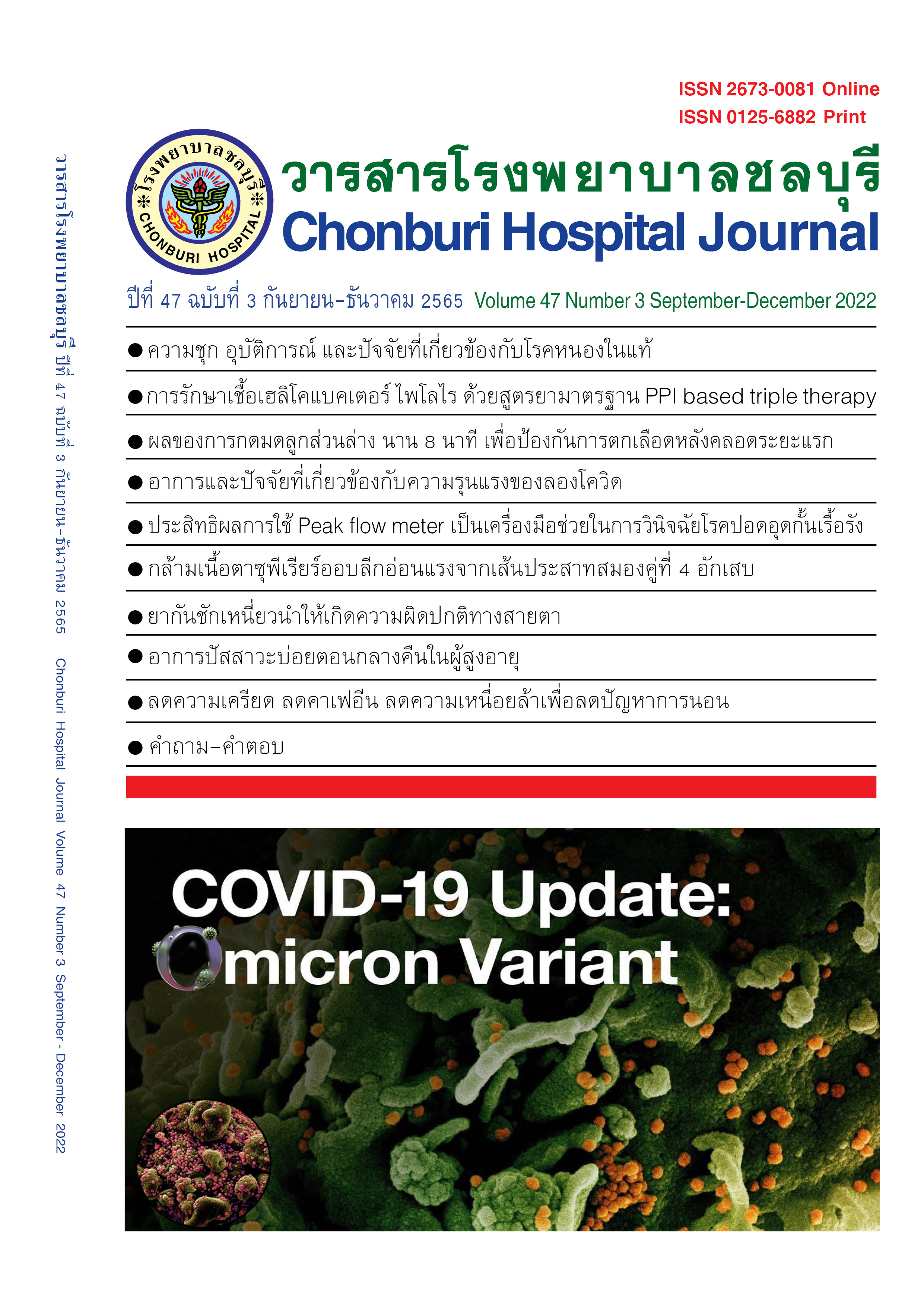
การศึกษาประสิทธิผลการใช้ Peak flow meter ร่วมกับแบบสอบถามและภาพรังสีทรวงอกเป็นเครื่องมือช่วยในการวินิจฉัยโรคปอดอุดกั้นเรื้อรังในเวชปฏิบัติ
บทคัดย่อ
บทนำ: การวินิจฉัยโรคปอดอุดกั้นเรื้อรังที่ถูกต้องมีความสำคัญต่อการรักษา ตามเกณฑ์การวินิจฉัยโรคปอดอุดกั้นเรื้อรังจำเป็นต้องอาศัยผลการตรวจ Spirometry ในประเทศไทยมีข้อจำกัดในการตรวจ Spirometry เนื่องจากการตรวจดังกล่าวไม่สามารถทำได้ในทุกโรงพยาบาล แต่การตรวจ Peak expiratory flow rate (PEFR) ด้วยเครื่อง Peak flow meter สามารถทำได้ง่ายกว่าและทำได้ทุกโรงพยาบาล งานวิจัยนี้จึงมีวัตถุประสงค์เพื่อศึกษาประสิทธิผลการใช้ Peak flow meter ร่วมกับแบบสอบถามและภาพรังสีทรวงอกเป็นเครื่องมือช่วยในการวินิจฉัยโรคปอดอุดกั้นเรื้อรัง วัสดุและวิธีการศึกษา: งานวิจัยนี้มีรูปแบบเป็น Cross sectional analytical study โดยผู้เข้าร่วมวิจัยจะได้รับการประเมินโดยแบบสอบถาม ได้รับการตรวจ PEFR Spirometry และภาพรังสีทรวงอก
ผลการศึกษา: มีผู้เข้าร่วมโครงการวิจัยจำนวน 196 คน ได้รับคัดเลือกให้เข้าร่วมวิจัย 141 คน เป็นโรคปอดอุดกั้นเรื้อรัง 39 คน (27.66%) โรคหืด 100 คน (70.93%) โรคหลอดลมพอง 2 คน (1.42%) ประวัติมีอาการหอบเหนื่อย (mMRC dyspnea scale ≥1) หรือไอหรือมีเสมหะเรื้อรังอย่างน้อย 6 เดือน มีความไว 100.0% ความจำเพาะ 3.9% ค่าพยากรณ์บวก 28.5% ค่าพยากรณ์ลบ 100.0% ค่า PEFR <80% predicted มีความไว 97.4% ความจำเพาะ 53.9% ค่าพยากรณ์บวก 44.7% ค่าพยากรณ์ลบ 98.2% ภาพรังสีทรวงอกปกติหรือมี hyperaeration มีความไว 69.2% ความจำเพาะ 5.9% ค่าพยากรณ์บวก 22.0% ค่าพยากรณ์ลบ 33.3% ในการวินิจฉัยโรคปอดอุดกั้นเรื้อรัง Area under the receiver operating characteristic curve (AUC) ในการวินิจฉัยโรคปอดอุดกั้นเรื้อรัง เมื่อทำนายด้วยปัจจัย อายุ เพศ ประวัติมีอาการหอบเหนื่อยหรือไอหรือมีเสมหะเรื้อรังอย่างน้อย 6 เดือน อายุที่เริ่มมีอาการ ≥40ปี ประวัติการสูบบุหรี่ การสัมผัสฝุ่นควันเป็นประจำ ประวัติวัณโรคปอดในอดีต PEFR <80% predicted และภาพรังสีทรวงอกปกติหรือมี hyperaeration ร่วมกัน ได้ AUC 96.67% (94.07 – 99.27)
สรุป: การใช้ข้อมูลประวัติจากแบบสอบถามร่วมกับค่า PEFR <80% predicted และภาพรังสีทรวงอกปกติหรือมี hyperaeration เป็นเครื่องมือที่มีประสิทธิภาพในการช่วยวินิจฉัยโรคปอดอุดกั้นเรื้อรัง
เอกสารอ้างอิง
World Health Organization. World Health Report [Internet]. Geneva: WHO; 2000 [cited 2018 Jan 23]. Available from: http://www.who.int/whr/2000/en/statistics.htm
National Health Security Office. Report of asthma and COPD project outcomes in 2012. [cited 2018 Jan 23]. Available from: http://www.nhso.go.th/frontend/NewsInformation Detail.aspr?newsld=Njgy
Global Initiative for Chronic Obstructive Lung Disease. Global strategy for the diagnosis, management, and prevention of chronic obstructive pulmonary disease (2018 report). n.p.: Global Initiative for Chronic Obstructive Lung Disease; 2018.
Tiyaworanan K. Spirometry test results and factors associated with quality of spirometry test in patients with chronic obstructive pulmonary disease (COPD) at Sisaket hospital. Srinagarind Med J 2016;31(6):348-54.
Sichletidis L, Spyratos D, Papaioannou M, Chloros D, Tsiotsios A, Tsagaraki V, et al. A combination of the IPAG questionnaire and PiKo-6® flow meter is a valuable screening tool for COPD in the primary care setting. Prim Care Respir J 2011;20(2):184-9.
Fritha P, Crockettb A, Beilbyc J, Marshalld D, Attewelle R, Ratnanesanf A, et al. Simplified COPD screening: validation of the PiKo-6® in primary care. Prim Care Respir J 2011;20(2):190-8.
Ching SM, Pang YK, Price D, Cheong AT, Lee PY, Irmi I, et al. Detection of airflow limitation using a handheld spirometer in a primary care setting. Respirology 2014;19:689–93.
Kobayashi S, Hanagama M, Yanai M. Early detection of chronic obstructive pulmonary disease in primary care. Intern Med 2017;56:3153-8.
Thorn J, Tilling B, Lisspers K, Jorgensen L, Stenling A, Stratelis G. Improved prediction of COPD in at-risk patients using lung function pre-screening in primary care: a real-life study and cost-effectiveness analysis. Prim Care Respir J 2012;21(2):159-66.
Kjeldgaard P, Lykkegaard J, Spillemose H, Ulrik CS. Multicenter study of the COPD-6 screening device: feasible for early detection of chronic obstructive pulmonary disease in primary care? Int J Chron Obstruct Pulmon Dis 2017;12:2323-31.
Jackson H, Hubbard R. Detecting chronic obstructive pulmonary disease using peak flow rate: cross sectional survey. Br Med J 2003;327:653-4.
Thorat YT, Salvi SS, Kodgule RR. Peak flow meter with a questionnaire and mini-spirometer to help detect asthma and COPD in real-life clinical practice: a cross sectional study. NPJ Prim Care Respir Med 2017;27(1):32. doi: 10.1038/s41533-017-0036-8.
Price DB, Tinkelman DG, Halbert RJ, Nordyke RJ, Isonaka S, Nonikov D, et al. Symptom-based questionnaire for identifying COPD in smokers. Respiration 2006;73:285-95.
Price DB, Tinkelman DG, Nordyke RJ, Isonaka S, Halbert RJ; for the COPD
Questionnaire Study Group. Scoring system and clinical application of COPD
Diagnostic Questionnaires. Chest 2006;129:1531-9.
Dejsomritrutai W, Nana A, Maranetra KN, Chuaychoo B, Maneechotesuwan K, Wongsurakiat P, et al. Reference spirometric values for healthy lifetime nonsmokers in Thailand. J Med Assoc Thai 2000;83(5):457-66.
Miller MR, Hankinson J, Brusasco V, Brugos F, Casaburi R, Coates A, et al. Standardisation of spirometry. Eur Respir J 2005;26(2):319-38. doi: 10.1183/09031936.05.00034805
ดาวน์โหลด
เผยแพร่แล้ว
เวอร์ชัน
- 2023-01-05 (2)
- 2022-12-28 (1)
ฉบับ
บท
การอนุญาต

This work is licensed under a Creative Commons Attribution-NonCommercial-NoDerivatives 4.0 International License.
บทความที่ได้รับการตีพิมพิ์เป็นลิขสิทธิ์ของวารสารโรงพยาบาลชลบุรี
