THE STUDY OF CHEST RADIOGRAPH OF POST COVID-19 PNEUMONIA AT RANONG HOSPITAL
Keywords:
COVID-19 pneumonia, Chest radiograph, Follow-up chest radiographAbstract
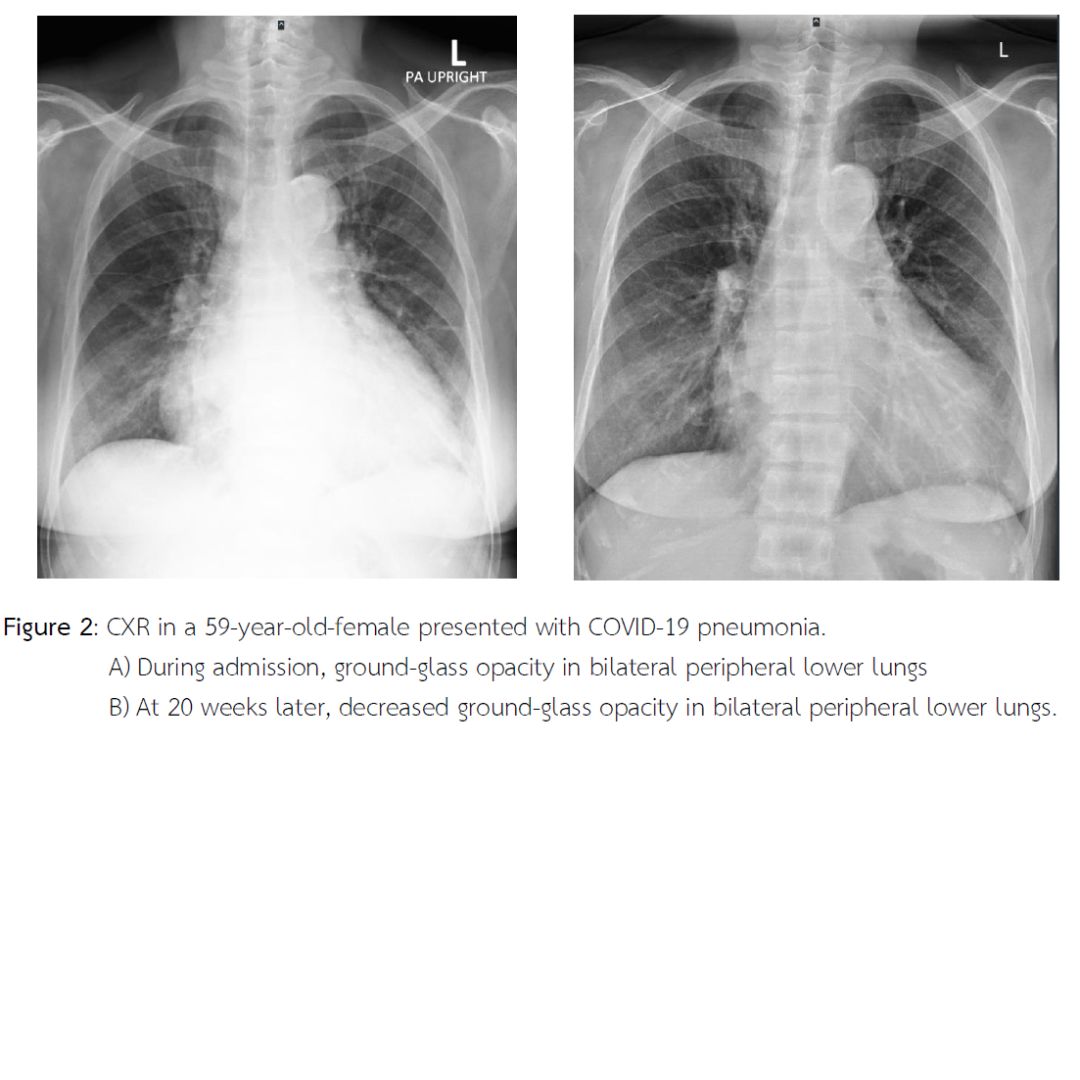
This research aimed to study the characteristics of chest radiographs following recovery from COVID-19 pneumonia in patients at Ranong Hospital between January 1 and December 31, 2022. Data collection was conducted using patient records and the Picture Archiving and Communication System (PACS). Descriptive statistics, including frequency distribution, percentage, mean, and standard deviation, were employed to describe general patient information and chest radiograph findings. The study included 166 patients with an average age of 66.8 years, comprising 51.2% females and 48.8% males. The average time between hospital admission and follow-up chest radiographs was 11.7 weeks. The results showed that 43 patients (25.9%) had completely normal chest radiographs, and 41 patients (24.7%) exhibited reduced abnormalities compared to initial imaging. Among the residual abnormalities, ground-glass opacity was observed in 43 patients (47.3%), consolidation in 31 patients (34%), reticular infiltration in 10 patients (11.0%), and other findings in 7 patients (7.7%). The residual abnormalities were predominantly distributed in the peripheral and lower lung regions bilaterally. In conclusion, the majority of post-recovery chest radiographs in COVID-19 pneumonia patients returned to normal, with only a subset of patients exhibiting residual abnormalities that could be detected via chest radiography. Therefore, chest radiography, which is widely available at all levels of healthcare in Thailand, is cost-effective and exposes patients to lower radiation doses compared to computed tomography (CT). It is an effective tool for initial follow-up of complications in patients recovering from COVID-19 pneumonia.
References
Alarcón-Rodríguez J. et al. (2021). Manejo y seguimiento radiológico del paciente post-COVID-19. Radiología, 63(3), 258–269. https://doi.org/10.1016/j.rx.2021.02.003
American College of Radiology. (2020). ACR recommendations for the use of chest radiography and computed tomography (CT) for suspected COVID-19 infection. https://www.acr.org/Advocacy-and-Economics/ACR-Position-Statements/Recommendations-for-Chest-Radiography-and-CT-for-Suspected-COVID19-Infection
British Thoracic Society. (2020). British Thoracic Society guidance on respiratory follow up of patients with a clinico-radiological diagnosis of COVID-19 pneumonia (Version 1.2). https://www.brit-thoracic.org.uk/covid-19/covid-19-information-for-the-respiratory-community/
Creamer A. W. et al. (2021). Clinico-radiological recovery following severe COVID-19 pneumonia. Thorax, 76(Suppl 1), A185. https://doi.org/10.1136/thorax-2020-BTSabstracts.320
Han X. et al. (2021). Six-month follow-up chest CT findings after severe COVID-19 pneumonia. Radiology, 299(1), E177–E186. https://doi.org/10.1148/radiol.2021203153
Lu H. et al. (2020). Outbreak of pneumonia of unknown etiology in Wuhan, China: The mystery and the miracle. Journal of Medical Virology, 92(4), 401–402. https://doi.org/10.1002/jmv.25678
Musat C. A. et al. (2021). Observational study of clinico-radiological follow-up of COVID-19 pneumonia: A district general hospital experience in the UK. BMC Infectious Diseases, 21(1), 1233. https://doi.org/10.1186/s12879-021-06941-8
National Institutes of Health of the U.S. Department of Health and Human Services. (2021). Coronavirus Disease 2019 (COVID-19) Treatment Guidelines. https://www.covid19treatmentguidelines.nih.gov/
Ojo A. S. et al. (2020). Pulmonary fibrosis in COVID-19 survivors: Predictive factors and risk reduction strategies. Pulmonary Medicine, 2020, 6175964. https://doi.org/10.1155/2020/6175964
Parry A. H. et al. (2021). Medium-term chest computed tomography (CT) follow-up of COVID-19 pneumonia patients after recovery to assess the rate of resolution and determine the potential predictors of persistent lung changes. The Egyptian Journal of Radiology and Nuclear Medicine, 52(1), 55. https://doi.org/10.1186/s43055-021-00434-z
Smith D. et al. (2020). A characteristic chest radiographic pattern in the setting of the COVID-19 pandemic. Radiology: Cardiothoracic Imaging, 2(5), e200280. https://doi.org/10.1148/ryct.2020200280

Downloads
Published
How to Cite
Issue
Section
License
Copyright (c) 2024 Primary Health Care Journal (Northeastern Edition)

This work is licensed under a Creative Commons Attribution-NonCommercial-NoDerivatives 4.0 International License.


