การศึกษาลักษณะภาพรังสีทรวงอกหลังรักษาหายของผู้ป่วยโรคปอดอักเสบจากโควิด 19 ในโรงพยาบาลระนอง
คำสำคัญ:
โรคปอดอักเสบจากโควิด 19, ภาพรังสีทรวงอก, การติดตามภาพรังสีทรวงอกบทคัดย่อ
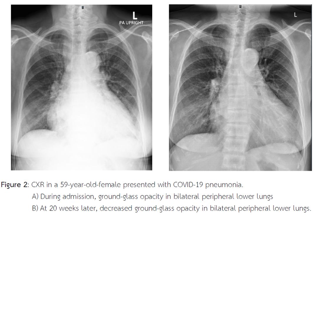
การศึกษาวิจัยนี้ มีวัตถุประสงค์เพื่อศึกษาลักษณะภาพรังสีทรวงอกหลังรักษาหายของผู้ป่วยโรคปอดอักเสบจากโควิด 19 ในโรงพยาบาลระนอง ระหว่างวันที่ 1 มกราคม ถึง 31 ธันวาคม พ.ศ.2565 โดยใช้เครื่องมือแบบบันทึกข้อมูลผู้ป่วยเป็นการสรุปข้อมูลผู้ป่วย เก็บรวบรวมข้อมูลจากเวชระเบียนและระบบ Picture archiving and communication system (PACS) และใช้สถิติเชิงพรรณนา ได้แก่ การแจกแจงความถี่ ร้อยละ ค่าเฉลี่ย และค่าเบี่ยงเบนมาตรฐาน สำหรับอธิบายข้อมูลทั่วไปทั้งข้อมูลส่วนส่วนบุคคลและข้อมูลลักษณะภาพทรวงอก ผลการศึกษาพบว่า กลุ่มตัวอย่างจำนวน 166 คน มีอายุเฉลี่ย 66.8 ปี เป็นเพศหญิงและเพศชายร้อยละ 51.2 และ 48.8 ตามลำดับ ระยะเวลาที่ถ่ายภาพรังสีทรวงอกนับจากวันที่นอนโรงพยาบาลและวันที่รักษาหายมีค่าเฉลี่ย 11.7 สัปดาห์ โดยพบว่าภาพรังสีทรวงอกเคยมีรอยโรคในวันที่นอนโรงพยาบาลหายเป็นปกติ และมีการลดลงของรอยโรค เท่ากับ 43 ราย (ร้อยละ 25.9) และ 41 ราย (ร้อยละ 24.7) ตามลำดับ ภาพรังสีทรวงอกที่ยังพบรอยโรคส่วนใหญ่เป็นลักษณะฝ้าขาว (Ground-glass opacity) และหนาทึบ เท่ากับ 43 ราย (ร้อยละ 47.3) และ 31 ราย (ร้อยละ 34) ตามลำดับ มีเพียงบางส่วนที่พบลักษณะเส้น (Reticular infiltration) และอื่น ๆ เท่ากับ 10 ราย (ร้อยละ 11) และ 7 ราย (ร้อยละ 7.7) ตามลำดับ พบลักษณะการกระจายของรอยโรคที่หลงเหลือเด่นที่ปอดส่วนนอกและส่วนล่าง (Peripheral, bilateral lower lungs) สรุป ส่วนใหญ่ภาพรังสีทรวงอกหลังรักษาหายของผู้ป่วยโรคปอดอักเสบจากโควิด 19 รอยโรคจะหายเป็นปกติ มีเพียงบางส่วนที่ยังพบรอยโรคและสามารถตรวจพบรอยโรคนั้นได้โดยใช้ภาพรังสีทรวงอก ดังนั้น ภาพรังสีทรวงอกที่มีพร้อมใช้ในสถานพยาบาลทุกระดับของประเทศไทย มีค่าใช้จ่ายที่น้อยกว่าและผู้ป่วยได้รับปริมาณรังสีน้อยกว่า เมื่อเทียบกับภาพเอกซเรย์คอมพิวเตอร์จึงเป็นเครื่องมือที่มีประสิทธิภาพเพียงพอในการเริ่มต้นติดตามภาวะแทรกซ้อนของผู้ป่วยปอดอักเสบจากโควิด 19
เอกสารอ้างอิง
Alarcón-Rodríguez J. et al. (2021). Manejo y seguimiento radiológico del paciente post-COVID-19. Radiología, 63(3), 258–269. https://doi.org/10.1016/j.rx.2021.02.003
American College of Radiology. (2020). ACR recommendations for the use of chest radiography and computed tomography (CT) for suspected COVID-19 infection. https://www.acr.org/Advocacy-and-Economics/ACR-Position-Statements/Recommendations-for-Chest-Radiography-and-CT-for-Suspected-COVID19-Infection
British Thoracic Society. (2020). British Thoracic Society guidance on respiratory follow up of patients with a clinico-radiological diagnosis of COVID-19 pneumonia (Version 1.2). https://www.brit-thoracic.org.uk/covid-19/covid-19-information-for-the-respiratory-community/
Creamer A. W. et al. (2021). Clinico-radiological recovery following severe COVID-19 pneumonia. Thorax, 76(Suppl 1), A185. https://doi.org/10.1136/thorax-2020-BTSabstracts.320
Han X. et al. (2021). Six-month follow-up chest CT findings after severe COVID-19 pneumonia. Radiology, 299(1), E177–E186. https://doi.org/10.1148/radiol.2021203153
Lu H. et al. (2020). Outbreak of pneumonia of unknown etiology in Wuhan, China: The mystery and the miracle. Journal of Medical Virology, 92(4), 401–402. https://doi.org/10.1002/jmv.25678
Musat C. A. et al. (2021). Observational study of clinico-radiological follow-up of COVID-19 pneumonia: A district general hospital experience in the UK. BMC Infectious Diseases, 21(1), 1233. https://doi.org/10.1186/s12879-021-06941-8
National Institutes of Health of the U.S. Department of Health and Human Services. (2021). Coronavirus Disease 2019 (COVID-19) Treatment Guidelines. https://www.covid19treatmentguidelines.nih.gov/
Ojo A. S. et al. (2020). Pulmonary fibrosis in COVID-19 survivors: Predictive factors and risk reduction strategies. Pulmonary Medicine, 2020, 6175964. https://doi.org/10.1155/2020/6175964
Parry A. H. et al. (2021). Medium-term chest computed tomography (CT) follow-up of COVID-19 pneumonia patients after recovery to assess the rate of resolution and determine the potential predictors of persistent lung changes. The Egyptian Journal of Radiology and Nuclear Medicine, 52(1), 55. https://doi.org/10.1186/s43055-021-00434-z
Smith D. et al. (2020). A characteristic chest radiographic pattern in the setting of the COVID-19 pandemic. Radiology: Cardiothoracic Imaging, 2(5), e200280. https://doi.org/10.1148/ryct.2020200280

ดาวน์โหลด
เผยแพร่แล้ว
วิธีการอ้างอิง
ฉบับ
บท
การอนุญาต
ลิขสิทธิ์ (c) 2024 วารสารสาธารณสุขมูลฐาน (ภาคตะวันออกเฉียงเหนือ)

This work is licensed under a Creative Commons Attribution-NonCommercial-NoDerivatives 4.0 International License.


