
External Fixation Device for Magnetic Resonance Imaging (MRI), Ramathibodi Hospital
Keywords:
radiographic studies, innovation, experimental development research, MRI, R2RAbstract
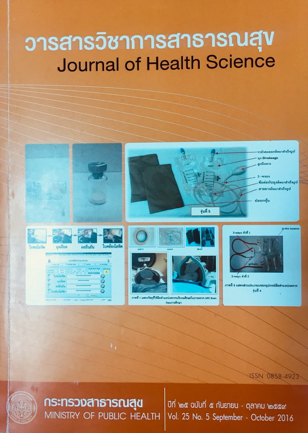
Magnetic Resonance Imaging (MRI) can accurately define various tissue and does not expose patients to harmful radiographic waves. In the process, the patient must lie relatively still for at least 30 minutes for an MRI. This can lead to patient fatigue and a feeling of discomfort. If the patient does not lie still, the image can be blurry or mal-positioned. The objective of this study was to develop an external fixation device that could be used in conjunction with MRI equipment to stabilize the body area being imaged. The study process was conducted from June 2013 to April 2015 with a total of 7,540 imaging studies completed (including head, neck, shoulder, wrist, knee, ankle, foot, etc). The control group consisted of 7,540 imaging studies and the intervention group consisted of 500 imaging studies. The product was produced from recycled materials; and the design was evaluated every 100 studies with a total of 5 revisions. Data were analyzed by descriptive statistics. It was found that the time necessary for an imaging study (outside of study time, which includes repositioning of patient for optimal image resolution) decreased from 40 minutes initially down to 20, 10 and 0 minutes with each revision of the product. The use of duck-tape reduced by an average of over 80 rolls per year. Furthermore, the device allowed the service system to improve MRI scheduling of patients. The device could be adjusted for various body parts, easy and convenient to use, and simple to maintain. It further decreased the technician’s labor in preparing patients for MRI. Healthcare providers and patients were satisfied with the device. Lastly, the cost of production was less than 500 baht. The authors recommend that every hospital should produce the device to be used in the MRI services.
Downloads
Downloads
Published
How to Cite
Issue
Section
License
Copyright (c) 2017 Journal of Health Science- วารสารวิชาการสาธารณสุข

This work is licensed under a Creative Commons Attribution-NonCommercial-NoDerivatives 4.0 International License.


